|
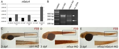
Evolutionarily conserved inhibition of nfatc4 by nfixa and regulation of primary slow fibers. (A) Quantitative real-time PCR (qRT-PCR) of nfatc4 mRNA expression normalized to ef1a. The expression of nfatc4 remains elevated in 48 hpf, 3 and 5 dpf nfixa-MO injected embryos, compared with controls. (B) RT-PCR on control and splice-nfatc4-MO-injected embryos at 24 hpf. RT-PCR primers were designed in exon 1 and exon 3, respectively; the amplification product, which contains the second exon, was 326 bp in control embryos, whereas two bands at 326 bp (arrowhead) and 174 bp (arrow) were detected in splice-nfatc4-MO injected embryos, confirming the partial skipping of the second exon (150 bp). (C-E) Immunostaining with F59 antibody (slow fibers): at 3 dpf, nfixa-nfatc4-MO injected embryos (E) present a F59 signal comparable with controls (C), whereas F59 remains elevated in nfixa-MO-injected embryos (D).
|