
FIGURE
Fig. 3
- ID
- ZDB-FIG-130108-3
- Publication
- Uribe et al., 2012 - Id2a functions to limit Notch pathway activity and thereby influence the transition from proliferation to differentiation of retinoblasts during zebrafish retinogenesis
- Other Figures
- All Figure Page
- Back to All Figure Page
Fig. 3
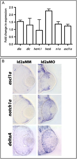
Notch pathway component gene expression is upregulated in Id2a-deficient retinae. (A) qRT-PCR quantification of 48 hpf dla, dlc, her4.1, hes6, n1a and ascl1a levels in Id2aMO and Id2aMM retinae. Transcript levels were normalized to tubulin, alpha 1 and the fold-change in expression in Id2aMO vs. Id2aMM presented. Error bars represent SEM, **p<0.05, n=3 biological replicates. (B) Transverse sections reveal the expression domains for ascl1a, notch1a and deltaA in Id2aMM and Id2aMO embryos at 48 hpf following in situ hybridization (n=6 sectioned embryos/condition). |
Expression Data
| Genes: | |
|---|---|
| Fish: | |
| Knockdown Reagent: | |
| Anatomical Terms: | |
| Stage: | Long-pec |
Expression Detail
Antibody Labeling
Phenotype Data
| Fish: | |
|---|---|
| Knockdown Reagent: | |
| Observed In: | |
| Stage: | Long-pec |
Phenotype Detail
Acknowledgments
This image is the copyrighted work of the attributed author or publisher, and
ZFIN has permission only to display this image to its users.
Additional permissions should be obtained from the applicable author or publisher of the image.
Reprinted from Developmental Biology, 371(2), Uribe, R.A., Kwon, T., Marcotte, E.M., and Gross, J.M., Id2a functions to limit Notch pathway activity and thereby influence the transition from proliferation to differentiation of retinoblasts during zebrafish retinogenesis, 280-292, Copyright (2012) with permission from Elsevier. Full text @ Dev. Biol.