
Fig. 4
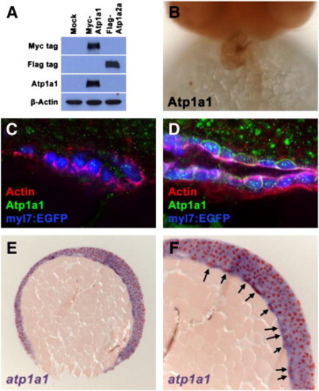
Endogenous Atp1a1 protein is not detected in the heart at stages when morphogenesis defects are first observed in atp1a1 mutants. (A) Western blot visualizing Myc-tagged, Flag-tagged, Atp1a1, and β-Actin proteins in HEK293T cell lysates. Mock transfected cell lysates have no reactivity with Myc, Flag, or Atp1a1 antibodies. Lysates from cells transfected with a Myc-tagged zebrafish Atp1a1 construct contain a protein that reacts with both Myc and Atpa1a antibodies. Lysates from cells transfected with a Flag-tagged zebrafish Atp1a2a construct contain a protein that reacts only with the Flag antibody. The presence of β-Actin protein in mock, Myc-Atp1a1, and Flag-Atp1a2a lysates demonstrates equal loading of total protein. (B) Ventral view of Atp1a1 antibody staining on 48 hpf wild type embryo. Atp1a1 protein is strongly detected in the heart. (C,D) Transverse vibratome sections of 20S stage (C) and 24 hpf (D) wild type embryos. At the 20S stage (C), Actin (red) is primarily basolateral around the cardiomyocytes of the cardiac cone (blue), whereas Atp1a1 protein (green) is not detectable in the cardiomyocyte membranes. By 24 hpf (D), polymerized Actin (red) is present on the ventral and lumenal surfaces of the primitive heart tube. Atp1a1 protein (green) is strongly localized to the membrane of cardiomyocytes in a punctate manner by this stage. (E,F) Microtome section of atp1a1 expression in 90% epiboly wild type embryo. (E) At 90% epiboly, atp1a1 is expressed ubiquitously. (F) Magnified portion of image in (E) shows that yolk syncytial layer nuclei strongly express atp1a1 (arrows). |
| Gene: | |
|---|---|
| Antibody: | |
| Fish: | |
| Anatomical Terms: | |
| Stage Range: | 90%-epiboly to Long-pec |
Reprinted from Developmental Biology, 362(2), Langenbacher, A.D., Huang, J., Chen, Y., and Chen, J.N., Sodium pump activity in the yolk syncytial layer regulates zebrafish heart tube morphogenesis, 263-270, Copyright (2012) with permission from Elsevier. Full text @ Dev. Biol.