
Fig. 3
- ID
- ZDB-FIG-111128-73
- Publication
- Muto et al., 2011 - Multifactorial Origins of Heart and Gut Defects in nipbl-Deficient Zebrafish, a Model of Cornelia de Lange Syndrome
- Other Figures
- All Figure Page
- Back to All Figure Page
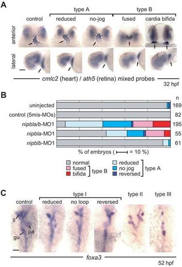
Effects of nipbl-MOs on heart and gut development. (A) Heart morphology was assessed at 32 hpf by cmlc2 expression. Head-heart regions of control embryos (leftmost panels) and nipbla/b-morphants are shown in anterior (upper panels) and lateral (lower panels) views. Type A embryos form heart tubes (dashed white lines) but jogging to the left is incomplete (“reduced”) or absent (“no-jog”). Type B embryos fail to form heart tubes, and fusion of cardiac precursors at the midline is partial (“fused”) or entirely lacking (“cardia bifida”). Simultaneous detection of ath5 expression throughout the dorsal and temporal retina in all embryos indicates no significant developmental delay in nipbla/b-morphants. Arrows point to heart tube or cardiac precursors. (B) Frequencies of different heart phenotypes in morphants; numbers (n) of embryos are on the right. (C) Gut and visceral organ morphology was assessed at 52 hpf by foxa3 expression. Dorsal views, with anterior to the top, are shown for control embryos (leftmost panel) and nipbla/b-morphants. Type I embryos have thin gut tubes as well as small liver and pancreas. Looping of the gut tube is also reduced, absent (no loop), or reversed. In type II embryos, anterior gut tubes are split, and associated visceral organs are bilaterally duplicated. Type III embryos have few or no cells expressing foxa3, and lack a gut tube. Scale bars: 50 μm. |
| Fish: | |
|---|---|
| Knockdown Reagents: | |
| Observed In: | |
| Stage Range: | Prim-15 to Long-pec |