
Fig. 5
- ID
- ZDB-FIG-111108-10
- Publication
- Hale et al., 2011 - Netrin Signaling Breaks the Equivalence between Two Identified Zebrafish Motoneurons Revealing a New Role of Intermediate Targets
- Other Figures
- All Figure Page
- Back to All Figure Page
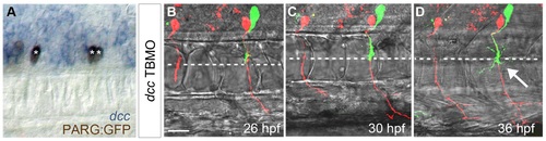
Dcc is necessary to prevent a second CaP/VaP axon from extending beyond the muscle pioneers. A. Brightfield image of lateral view of 24 hpf embryo focusing on the spinal cord. dcc mRNA (blue) is co-localized with a motoneuron marker, PARG:GFP (brown; see also [47]). The left segment shows one CaP (*) expressing dcc. The right segment shows two CaP/VaPs (**) expressing dcc. dcc is also expressed in additional cells that are likely other motoneurons and neighboring interneurons. B-D. Projected confocal stacks of living dye-labeled motoneurons in the same dcc TBMO-injected embryo at three developmental stages. Punctate red and green fluorescence within the spinal cord but not in CaP or VaP in this and other figures represents dye leakage from the labeling procedure. The left segment shows a CaP (red) in a segment lacking VaP; the right segment shows both CaP (red) and VaP (green). B. At 26 hpf the VaP axon does not extend beyond the level of the muscle pioneers (dashed line) but CaP axons extend farther. C. By 30 hpf, CaP axons have extended farther ventrally but the VaP axon still remains near the muscle pioneers. D. By 36 hpf, CaP axons have reached the ventral aspect of the myotome, providing evidence that Dcc is unnecessary for CaP axon extension. The VaP axon has extended branches beyond and along the muscle pioneers (arrow). Scale bar = 20 µm. |
| Gene: | |
|---|---|
| Fish: | |
| Anatomical Terms: | |
| Stage: | Prim-5 |
| Fish: | |
|---|---|
| Knockdown Reagent: | |
| Observed In: | |
| Stage: | Prim-5 |