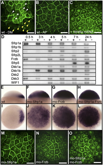
Negative regulation of the canonical Wnt pathway by Sfrp1a and Frzb. Immunofluorescence localization of β-catenin is shown at 3 hpf (high stage) (A), at the dorsal margin (DM), and (B) at the animal pole (AP) of a wild-type embryo or (C) at the animal pole of an embryo overexpressing Wnt8a (10 pg Wnt8a mRNA). White and yellow arrowheads in A point to the accumulation of β-catenin in blastomeres and yolk syncytial layer nuclei, respectively. (D) Analysis by RT-PCR of the expression of zebrafish Wnt inhibitors during early development. Sfrp (TLC), expressed only during gastrulation (35), is not presented here. Expression of chordin at the sphere stage in lateral view (Upper) and animal pole view (Lower) in (E and I) wild-type embryos, (F and J) Sfrp1a morphants, (G and K) Frzb morphants, and (H and L) Sfrp1a–Frzb double morphants. (M–O) Immunofluorescence localization of β-catenin in nuclei of the animal pole of embryos at the high stage in (J) Sfrp1a, (K) Frzb, and (L) Sfrp1a–Frzb morphants. Embryos are in dorsal view (A), lateral view (E–H), and in animal pole view (B, C, and I–O). (Scale bar, 100 μM.)
|
