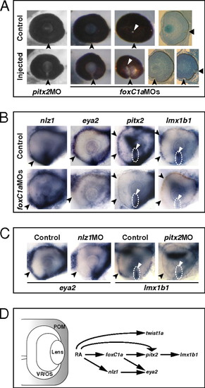
Abrogation of FoxC1a, Nlz1, and Pitx2 function causes coloboma and affects gene expression in POM. (A) Eyes of embryos injected with pitx2 MO or foxC1a MOs showing coloboma at 72 hpf, as detected in lateral view (Left and Center Left), posterior view (Center Right), and histological sections (Right). The injected eyes exhibit an open choroid fissure (arrowheads) ventral to the optic disk (triangles). (B and C) Lateral views of 32-hpf eyes. Panels are representative images of two to three independent experiments. Approximately 10–20 embryos were used for each condition and probe in each experiment. (B) Decreased expression of eya2, pitx2, and lmx1b1, but not nlz1, in choroid fissure POM (dashed circles and triangles) and/or anterior POM (arrowheads) in the foxC1a MO-injected embryos. (C) Decreased expression of eya2 in anterior POM (arrowheads) of an nlz1 MO-injected embryo and of lmx1b1 in anterior POM (arrowheads) and within the choroid fissure (dashed circles and triangles) of a pitx2 MO-injected embryo. (D) Proposed model of the molecular mechanisms of RAR-dependent RA signaling during development of the zebrafish POM (highlighted in gray). See text for details.
|
