
Fig. 2
- ID
- ZDB-FIG-110622-121
- Publication
- Lupo et al., 2011 - Retinoic acid receptor signaling regulates choroid fissure closure through independent mechanisms in the ventral optic cup and periocular mesenchyme
- Other Figures
- All Figure Page
- Back to All Figure Page
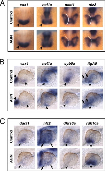
RAR signaling regulates gene expression in the VR/OS. (A–C) Embryos treated with DMSO or 10 μM AGN from 3s and hybridized at 18s (A) or 24–25 hpf (B and C) with the indicated probes. (A) Dorsal/anterior views of heads. AGN treatment has no major effects on the expression of vax1 and net1a at the optic vesicle/forebrain junction (arrowheads) and of dact1 and nlz2 in the optic vesicle. Dashed lines highlight the proximal edge of the optic vesicle. (B and C) Lateral views of eyes. AGN treatment causes down-regulation of vax1, net1a, cyb5a, itgA5, dact1, nlz2, and dhrs3a and up-regulation of rdh10a in the VR/OS (arrowheads). Triangles point to vax1 expression in the ventral forebrain (B). Arrows point to itgA5 expression in extraocular tissue at the back of the eye (B) and nlz2 expression on each side of the choroid fissure (C). Panels are representative images of the following numbers (n) of independent experiments: vax1, net1a, n = 4; itgA5, n = 3; and cyb5a, dact1, nlz2, dhrs3a, rdh10a, n = 2. Approximately 15–30 embryos were assayed for each condition and probe in each experiment. |
| Genes: | |
|---|---|
| Fish: | |
| Anatomical Terms: | |
| Stage Range: | 14-19 somites to Prim-5 |