|
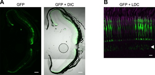
Expression of GFP in a Tg(LAR:LWS2up1.8kb:GFP)#1501 retina. (A) A transverse section of a Tg(LAR:LWS2up1.8kb:GFP)#1501 retina. The left panel shows the image of GFP signals (green) and the right panel shows the overlay with its DIC image. The dorsal side is oriented at the top of each panel and the ventral side is at the bottom. (B) A vertical and expanded view of the photoreceptor layer of the same retina as shown in (A). GFP (green) was specifically expressed in LDCs, whose outer segments were immunostained with the antibody against the zebrafish red opsin (magenta). Arrowheads indicate the faint GFP signals detected in some bipolar cells. Scale bars = 100 μm (A), 10 μm (B).
|