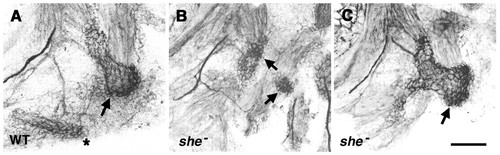
Two classes of opercle osteoblast clusters, expanded and reduced, are present in she mutants. Labeling with the zns5 monoclonal antibody in larvae at 88 hours postfertilization. Left-side views with dorsal to the top. The antibody labels both osteoblasts and nerve axons prominently, and muscles lightly. (A) wild type (WT). Osteoblasts envelop the opercle (arrow) and branchiostegal ray (asterisk). The dark lines are nerves. Delicately labeled muscles (at the top) connect separately to the opercle joint region (dilator operculi muscle, left) and blade region (adductor operculi, right). (B) A she mutant with a presumed mild opercle-loss phenotype. The branchiostegal ray osteoblast cluster is missing. Two clusters are present (arrows) that seem to correspond to the opercle joint region (upper, identified by its position and its connection with the dilator operculi muscle) and a diminished opercle `fan′ region (lower). A confocal z-series reveals no osteoblasts connect between these two clusters. (C) A she mutant with presumed opercle gain. The opercle osteoblast cluster is expanded (arrow), particularly at the joint region. The branchiostegal ray cluster is either missing or possibly is present but fused with the opercle cluster. Muscle connections are approximately normal. Scale bar: 50 μm.
|
