
FIGURE
Fig. 2
- ID
- ZDB-FIG-101123-37
- Publication
- Kim et al., 2010 - PDGF signaling is required for epicardial function and blood vessel formation in regenerating zebrafish hearts
- Other Figures
- All Figure Page
- Back to All Figure Page
Fig. 2
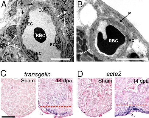
Mural cells are present in coronary vessels in the wound during zebrafish heart regeneration. (A and B) Transmission EM images of coronary vessels in the wound of 14-dpa regenerating hearts. EC, endothelial cells. P, pericytes. RBC, red blood cells. (Scale bar = 5 μm.) ISH using digoxigenin-labeled antisense probes against tagln (C) and acta2 (D) in sham-operated and 14-dpa regenerating hearts. The dashed red line marks the approximate position of the amputation plane. (Scale bar = 100 μm.) |
Expression Data
| Genes: | |
|---|---|
| Fish: | |
| Condition: | |
| Anatomical Term: | |
| Stage: | Adult |
Expression Detail
Antibody Labeling
Phenotype Data
Phenotype Detail
Acknowledgments
This image is the copyrighted work of the attributed author or publisher, and
ZFIN has permission only to display this image to its users.
Additional permissions should be obtained from the applicable author or publisher of the image.
Full text @ Proc. Natl. Acad. Sci. USA