
FIGURE
Fig. 4
- ID
- ZDB-FIG-100903-44
- Publication
- Wang et al., 2010 - Moesin1 and Ve-cadherin are required in endothelial cells during in vivo tubulogenesis
- Other Figures
- All Figure Page
- Back to All Figure Page
Fig. 4
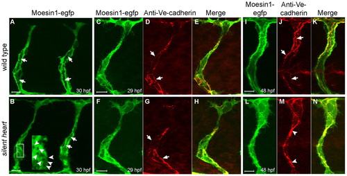
Primary endothelial lumen formation in the ISVs does not require blood flow. (A,B) Formation of the primary lumen (arrows) at 30 hpf is observed with the Tg(flk1:moesin1-egfp) line in either wild-type (A) or silent heart mutants (B). (B) silent heart embryos also displayed vacuoles during tubulogenesis (arrowheads in inset). (C-H) Ve-cadherin-labeled junctions (arrows) appeared normal at 29 hpf in silent heart mutants. (I-N) At 48 hpf, Ve-cadherin-labeled junctions (arrows) are often clustered in the silent heart embryos (arrowheads), which is likely to reflect the collapse of the primary lumen. |
Expression Data
| Genes: | |
|---|---|
| Antibody: | |
| Fish: | |
| Anatomical Terms: | |
| Stage Range: | Prim-15 to Long-pec |
Expression Detail
Antibody Labeling
Phenotype Data
| Fish: | |
|---|---|
| Observed In: | |
| Stage Range: | Prim-15 to Long-pec |
Phenotype Detail
Acknowledgments
This image is the copyrighted work of the attributed author or publisher, and
ZFIN has permission only to display this image to its users.
Additional permissions should be obtained from the applicable author or publisher of the image.
Full text @ Development