
FIGURE
Fig. 8
- ID
- ZDB-FIG-100616-99
- Publication
- Insinna et al., 2010 - Analysis of a zebrafish dync1h1 mutant reveals multiple functions for cytoplasmic dynein 1 during retinal photoreceptor development
- Other Figures
- All Figure Page
- Back to All Figure Page
Fig. 8
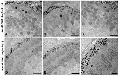
Knock-down of Dync1h1 phenocopies cnb mutants. (A-C) TEM of retinas from 3-dpf embryos injected with a high dose (8 ng/embryo) of dync1h1 morpholino (MO). Photoreceptors across the entire retina failed to form outer segments and had rounded nuclei (N). (D-L) TEM views of embryos injected with a lower amount (3 ng/embryo) of dyn1 hc1 morpholino. Outer segments formed in some photoreceptors at the central (D, E) and peripheral retina (F) (arrows). Most lower dose morphant cells still failed to elongate outer segments. Scale bars: 10 μm in (A-E); 2 μm in (F). |
Expression Data
Expression Detail
Antibody Labeling
Phenotype Data
| Fish: | |
|---|---|
| Knockdown Reagent: | |
| Observed In: | |
| Stage: | Protruding-mouth |
Phenotype Detail
Acknowledgments
This image is the copyrighted work of the attributed author or publisher, and
ZFIN has permission only to display this image to its users.
Additional permissions should be obtained from the applicable author or publisher of the image.
Full text @ Neural Dev.