
FIGURE
Fig. 4
- ID
- ZDB-FIG-100616-36
- Publication
- Boije et al., 2010 - Pax2 is expressed in a subpopulation of Müller cells in the central chick retina
- Other Figures
- All Figure Page
- Back to All Figure Page
Fig. 4
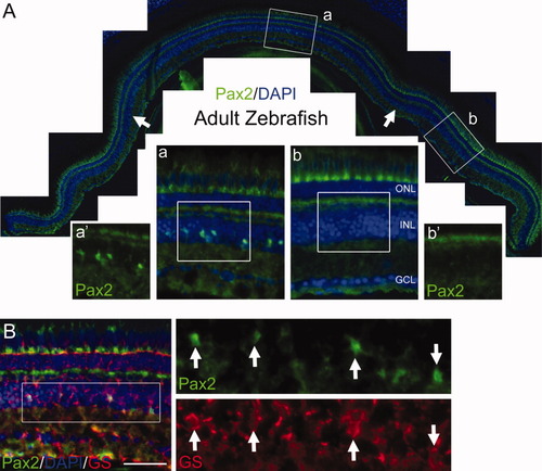
Pax2 labeling in the zebra fish retina. A: Epifluorescence micrograph from a whole section of adult retina showing Pax2 labeling. Arrows indicate the limits of the central Pax2 labeling. a,b: Magnifications of the boxed regions in A. a′,b′: The green channel of the boxed regions in (a) and (b). B: Pax2 co-labeling with the Müller cell marker glutamine synthetase (GS). Arrows indicate Pax2, GS double-positive cells. ONL, outer nuclear layer; INL, inner nuclear layer; GCL, ganglion cell layer. Scale bar = 25 μm in B (applies to (a) and (b)). |
Expression Data
| Antibodies: | |
|---|---|
| Fish: | |
| Anatomical Term: | |
| Stage: | Adult |
Expression Detail
Antibody Labeling
Phenotype Data
Phenotype Detail
Acknowledgments
This image is the copyrighted work of the attributed author or publisher, and
ZFIN has permission only to display this image to its users.
Additional permissions should be obtained from the applicable author or publisher of the image.
Full text @ Dev. Dyn.