
Fig. 2
- ID
- ZDB-FIG-100322-3
- Publication
- Tobin et al., 2010 - The lta4h Locus Modulates Susceptibility to Mycobacterial Infection in Zebrafish and Humans
- Other Figures
- All Figure Page
- Back to All Figure Page
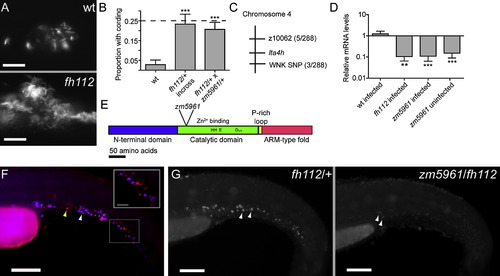
Mutations in the leukotriene A4 hydrolase Gene Result in Decreased lta4h mRNA Expression and Increased Susceptibility to Mycobacterial Infection (A) Fluorescence images of infection foci at 5 dpi with initial dose of 115 bacteria showing well-formed granuloma in WT fish (top) and profuse bacterial clusters showing cording morphology in hypersusceptible mutant fh112 sibling (bottom). The scale bar represents 25 μm. (B) Proportion of fish with cording morphology at 5 dpi with 150–250 bacteria in five independent clutches of 50–100 animals each of fh112/+ heterozygote or WT crosses and three independent clutches of 20–25 animals each from fh112/+ x zm5961/+ heterozygote crosses. Dotted line represents the theoretical maximum for a completely penetrant recessive mutation. Error bars represent the standard deviation (SD). ***p < 0.001 (one-way ANOVA with Tukey′s post-test). (C) Genetic map of fh112 placing it between the polymorphic markers z10062 and a SNP in a PRKWNK1-related gene on zebrafish chromosome 4. Parentheses indicate the number of recombination events as a ratio of the total number of informative meioses scored. (D) Mean lta4h RNA levels measured by qRT-PCR from 3–5 biological replicates of 30 fish. Infected fh112 mRNA assessed at 4 dpi, infected and uninfected zm5961 RNA at three dpf (1 dpi for infected zm5961 fish). Fold difference expressed relative to matched WT uninfected controls. **p < 0.01; ***p < 0.001 (ANOVA with Tukey′s post-test). Error bars represent the SD. (E) Gene structure of zebrafish lta4h with location of the zm5961 insertion and key conserved residues in the catalytic domain indicated (Haeggstrom, 2004). (F) Fluorescent in situ hybridization (FISH) of lta4h (red) combined with mpo antibody staining for neutrophils (blue) in caudal hematopoietic tissue of two dpf uninfected fish. Yellow arrowhead, lta4h-staining, mpo-negative presumed macrophage. White arrowhead, dual staining neutrophil. Scale bar, 100 μm. Inset scale bar represents 25 μm. (G) lta4h FISH in uninfected fh112/+ heterozygote (left) and zm5961/fh112 non-complementing sibling (right) at two dpf. Arrowheads point to brightly staining cells in fh112/+ heterozygote and to weakly-expressing cells in non-complementer. The scale bar represents 100 μm. See also Figure S1. |
| Gene: | |
|---|---|
| Fish: | |
| Anatomical Terms: | |
| Stage: | Long-pec |
Reprinted from Cell, 140(5), Tobin, D.M., Vary, J.C. Jr, Ray, J.P., Walsh, G.S., Dunstan, S.J., Bang, N.D., Hagge, D.A., Khadge, S., King, M.C., Hawn, T.R., Moens, C.B., and Ramakrishnan, L., The lta4h Locus Modulates Susceptibility to Mycobacterial Infection in Zebrafish and Humans, 717-730, Copyright (2010) with permission from Elsevier. Full text @ Cell