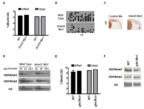
G9a and dnmt3 morphants have wild type 5-methyl-deoxycytosine and histone H3K9 di- or tri-methyl levels. (A) LC-MS assay was performed on genomic DNA isolated from dnmt3 morphants and wild type embryos at 24hpf and 72hpf. Note that no change in global 5-methyl-deoxycytosine levels was detected in dnmt3 morphants compared to wild type embryos at the time points tested. (B) Schematic presentation of bisulfite sequencing data for ntl CpG island in wild type and dnmt3 morphants at 24hpf. Black and white circles represent methylated and unmethylated cytosines in CpG sequences, respectively. (C) Expression of ntl (ventral view) in control and dnmt3 morphant embryos at 24hpf as detected by whole mount in situ hybridization. (D) Protein was isolated from wild type and dnmt3 morphants at 80hpf and H3K9me2 and H3K9me3 levels were measured by antibodies specific to these marks. Decreasing amounts (as shown in microgram quantities) of the proteins were loaded for proteins from dnmt3 morphants or wild type embryos. (E) LC-MS assay was performed on genomic DNA isolated from g9a morphants and wild type embryos at 24hpf and 72hpf. Note that no change in global 5-methyl-deoxycytosine levels was detected in g9a morphants compared to wild type embryos at the time points tested. (F) Protein was isolated from wild type and g9a morphants at 80hpf and H3K9me2 and H3K9me3 levels were measured by antibodies specific to these marks. Twenty five micrograms of total protein isolated from g9a morphants or wild type embryos was loaded in each lane. No detectable change was observed in global levels of H3K9me2 or H3K9me3 in dnmt3 or g9a morphants when compared to wild type embryos. Pan-H3 C-terminal antibody was used for total H3 content.
|
