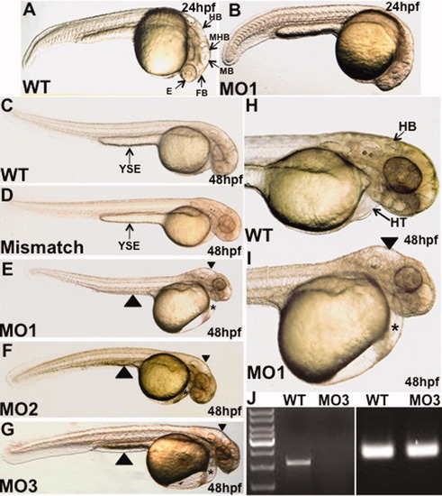
The mga morphants show defects in organogenesis of the brain, heart, and gut. Shown are representative control (wildtype: WT, or mismatch) or morphant (MO1, MO2, MO3) embryos as indicated. A,B: At 24 hpf, compared to control embryos, the morphant fails to partition normally the forebrain, midbrain, and hindbrain structures, and eye development is significantly delayed. C-I: At 48 hpf, mga morphants show a non-looping heart within a cardiac edema (asterisks), an edemic hindbrain cavity (arrowheads in E, F, G, and I), and a regression of the yolk stalk extension (large arrowheads in E-G), indicative of an endoderm defect. The morphant phenotype (brain, heart, and yolk stalk defects) shown here by representative embryos was documented for MO1 (100%, 320/320 embryos), MO2 (75%, 170/228 embryos), and MO3 (29%, 46/161 embryos). Views are lateral, dorsal to the top, and anterior to the right. HB, hindbrain; MHB, midbrain-hindbrain boundary; MB, midbrain; FB, forebrain; E, eye; HT, heart; YSE, yolk stalk extension. J: RT-PCR analysis demonstrates that the MO3 morpholino disrupts the production of normal transcript as shown by the absence of mga cDNA (left). The cDNA was prepared from control wildtype (WT) or morphant (MO3) embryos. Right panel shows equal amplification of β-actin for both WT and MO3 cDNA samples.
|
