
FIGURE
Fig. S2
Fig. S2
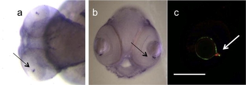
Mural cells in the inner optic circle of 72 hpf zebrafish larvae, (a and b) whole mount in situ hybridization with acta2 shows a specific staining in the inner optic circle (IOC) (black arrows), the major central retinal vessel that surrounds the lens, (c) immunofluorescence analysis of Tg(Flk1:GFP)s843 embryos with SM22alpha antibody (red) shows the presence and localization (white arrow) of MCs in the eye. Scale bar: 0.1 mm. |
Expression Data
| Genes: | |
|---|---|
| Antibody: | |
| Fish: | |
| Anatomical Terms: | |
| Stage: | Protruding-mouth |
Expression Detail
Antibody Labeling
Phenotype Data
Phenotype Detail
Acknowledgments
This image is the copyrighted work of the attributed author or publisher, and
ZFIN has permission only to display this image to its users.
Additional permissions should be obtained from the applicable author or publisher of the image.
Reprinted from Mechanisms of Development, 126(8-9), Santoro, M.M., Pesce, G., and Stainier, D.Y., Characterization of vascular mural cells during zebrafish development, 638-649, Copyright (2009) with permission from Elsevier. Full text @ Mech. Dev.