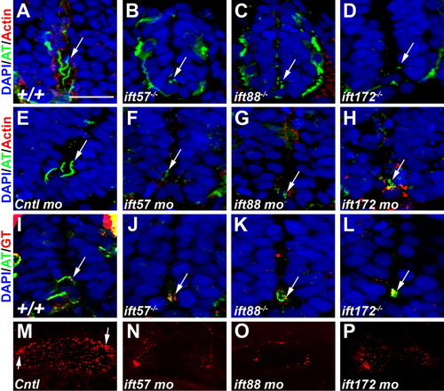
Loss of IFT disrupts ciliogenesis. A-D: Acetylated tubulin antibody (green) labeled primary cilia extending from cells in the ventral neural tube of 24-hpf wild type embryos. E-H: Cilia were disrupted in ift57, ift88, and ift172 mutant embryos at 24 hpf. Embryos injected with a control morpholino show cilia. Cilia were disrupted in 24-hpf embryos following injection of morpholinos against IFT genes. In morphant embryos, stabilized microtubules were still observed in small puncta that likely correspond to the basal body region. Arrows point to regions of acetylated tubulin staining. Rhodamine phalloidin (red) labels actin filaments. I-L: Gamma tubulin (red) recognized basal bodies that colocalized with acetylated tubulin (green) in the mutant embryos. In all sections nuclei are visualized using the nuclear stain DAPI. M-P: Fluorescent whole-mount images of the otic vesicle stained with acetylated tubulin at 18 hpf. Control embryos show clusters of longer cilia at the anterior and posterior edges (arrows) and numerous short cilia throughout the lumen. Cilia within the lumen are highly reduced or absent in IFT morphant embryos. The otic vesicles are also slightly smaller than control embryos. Scale bar = 20 μm for A-L.
|
