
Fig. 2
- ID
- ZDB-FIG-090504-10
- Publication
- Zeng et al., 2009 - Phospholipase D1 is required for angiogenesis of intersegmental blood vessels in zebrafish
- Other Figures
- All Figure Page
- Back to All Figure Page
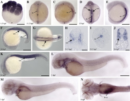
Spatio-temporal expression of pld1 during zebrafish embryogenesis. (A) pld1 transcripts are ubiquitously distributed at 16-cell stage (1.5 hpf). At 11.3 hpf (B, C) and 15 hpf (D, E), pld1 expression is restricted to the dorsal notochord (nc) and the adjacent forming somites (sm). (F–I) pld1 expression in somites (sm) becomes more pronounced during late segmentation stages. (H, I) Cross section of an embryo at 21.5 hpf shows pld1 expression in the somites (sm) in the anterior trunk region (H) but in notochord (nc) in the posterior trunk (I). (J, K) At 24 hpf, pld1 is primarily expressed in the somites (sm). (L) At 2 dpf, the expression of pld1 is strongly reduced in the somites and appears in the pharyngeal pouch (pp). (M, N) At 3 dpf and 4 dpf, pld1 is detected mainly in the liver. (A) Animal view: (B, D) dorsal views, animal pole towards the top; (C,E,F,K,L,M) lateral views, anterior to the left; (G, N) dorsal view, anterior to the left. Animal pole (☆, ★). Vegetal pole (#). Dorsal (D). Scale bar represents 100 μm (H, J) and 250 μm (A, C, E, F, K, L). |
| Gene: | |
|---|---|
| Fish: | |
| Anatomical Terms: | |
| Stage Range: | 16-cell to Day 4 |
Reprinted from Developmental Biology, 328(2), Zeng, X.X., Zheng, X., Xiang, Y., Cho, H.P., Jessen, J.R., Zhong, T.P., Solnica-Krezel, L., and Brown, H.A., Phospholipase D1 is required for angiogenesis of intersegmental blood vessels in zebrafish, 363-376, Copyright (2009) with permission from Elsevier. Full text @ Dev. Biol.