
Fig. 3
- ID
- ZDB-FIG-090310-7
- Publication
- Buckles et al., 2004 - Combinatorial Wnt control of zebrafish midbrain-hindbrain boundary formation
- Other Figures
- All Figure Page
- Back to All Figure Page
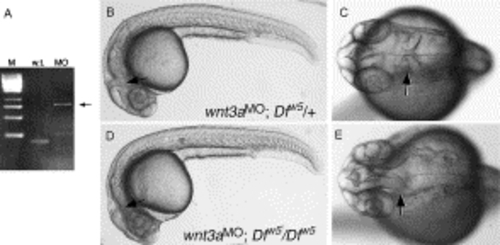
Loss of the isthmic constriction in embryos lacking Wnt3a, Wnt1 and Wnt10b. (A) RT-PCR analysis of wnt3a splice-blocking MO activity. The wild-type product (w.t.) is 168 bp. The morpholino blocks splicing of intron 1, leading to the predicted 810 bp PCR product (MO, arrow). (B–E) Live embryos at 27 hpf, anterior to the left. (B,C) Wild type phenotype observed in ∼75% of wnt3a MO injected embryos derived from a Dfw5/+intercross. The isthmic constriction (arrow) is easily seen in the dorsal view of the head in (C). (D,E) MHB-absent phenotype observed in ∼25% of injected embryos. Note the wild type appearance of the animal except for the loss of the isthmic constriction (arrows). |
| Fish: | |
|---|---|
| Knockdown Reagent: | |
| Observed In: | |
| Stage: | Prim-5 |
Reprinted from Mechanisms of Development, 121(5), Buckles, G.R., Thorpe, C.J., Ramel, M.C., and Lekven, A.C., Combinatorial Wnt control of zebrafish midbrain-hindbrain boundary formation, 437-447, Copyright (2004) with permission from Elsevier. Full text @ Mech. Dev.