
Fig. 2
- ID
- ZDB-FIG-090306-4
- Publication
- Sukumaran et al., 2009 - Early defects in photoreceptor outer segment morphogenesis in zebrafish ift57, ift88 and ift172 Intraflagellar Transport mutants
- Other Figures
- All Figure Page
- Back to All Figure Page
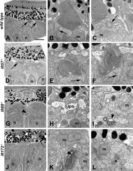
Transmission electron micrographs of transverse sections along the dorsal–ventral axis of wild type, ift57, ift88 and ift172 mutant zebrafish at 72 hpf. (A) Electron micrograph of a wild type embryo showed photoreceptor outer segments (arrows) that were easily observed at low magnification. (B and C) Higher magnification showed that outer segment disk membranes are well organized and the connecting cilium (arrows) can be seen. These cilia were approximately 300 nm in width and 500 nm in length. Mitochondria (m) were observed in the apical inner segment. (D) In ift57 mutants, outer segments were observed at low magnification. (E and F) At higher magnification, the disk membranes were well ordered but the outer segments appear somewhat disheveled compared to wild type. Arrows denote connecting cilia. (G) In ift88 mutants, no outer segments were seen and disorganized membrane structures (arrowhead) were seen near the nuclei (n). (H and I) Higher magnification images revealed the interphotoreceptor space (ips) between the RPE and the mitochondria (m), as well as membrane structure (arrowheads) located basal to the mitochondria. (J) Electron micrograph of an ift172 mutant also showed no outer segments. (K and L) Disorganized membrane structures (arrowhead) running parallel to the plasma membrane were seen basal to the mitochondria (m). A centriole (asterisk) that may be part of a mispositioned basal body was also seen several hundred nanometers away from the apical surface and below a mitochondria (m). Scale bar = 4 μm (A, D, G, and J), and 400 nm (B, C, E, F, H, I, K, and L). |
| Fish: | |
|---|---|
| Observed In: | |
| Stage: | Protruding-mouth |
Reprinted from Vision Research, 49(4), Sukumaran, S., and Perkins, B.D., Early defects in photoreceptor outer segment morphogenesis in zebrafish ift57, ift88 and ift172 Intraflagellar Transport mutants, 479-489, Copyright (2009) with permission from Elsevier. Full text @ Vision Res.