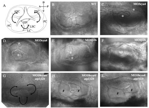
Otic vesicle morphology in wild-type and morphant fish (72 hpf). (A) Schematic of the normal otic vesicle structures visualized by differential interference contrast (DIC) imaging. Anterior, lateral and posterior cristae (AC, LC and PC, respectively) have differentiated into anterior, lateral and posterior semicircular canals (ASC, LSC and PSC, respectively) that are portioned by epithelial pillars (EP) inside the otic vesicle. (B) DIC image of the otic vesicle in wild-type fish reveals normal structures. (C,D) DIC images of otic vesicles from eya4 morphant fish show aborted protrusions of presumptive epithelial pillars (asterisks) into the vesicle and malformed canals. (E,F) DIC images of atp1b2b morphant fish otic vesicles show incomplete fusion and diminutive epithelial pillars (asterisks), which resemble those of the eya4 morphants. (G-I) atp1b2b mRNA-rescued eya4 morphant fish otic vesicles were still smaller than wild type, but were larger than eya4 morphant vesicles. Canal formation was partially (# in H,I) or nearly completely restored (G) as a result of the more mature epithelial pillars.
|
