
FIGURE
Fig. 5
- ID
- ZDB-FIG-080922-8
- Publication
- Guo et al., 1999 - Mutations in the zebrafish unmask shared regulatory pathways controlling the development of catecholaminergic neurons
- Other Figures
- All Figure Page
- Back to All Figure Page
Fig. 5
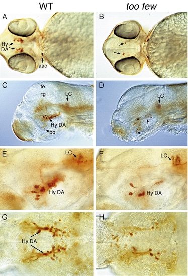
too few mutant embryos have reduced number of hypothalamic neurons. (A, B, and G, H) Ventral and (C–F) lateral views of wildtype (left) and too few mutant (right) embryos labeled with anti-TH antibody. (A–F) 2-day-old embryos showing reduced number of hypothalamic DA neurons but normal complement of postoptic DA and LC neurons. (G and H) 96-h fry showing that the reduction of hypothalamic DA neurons persist. Abbreviations: Hy DA, hypothalamic DA neurons; po, postoptic region; te, tectum, tg, tegmentum; Scale bar: 150 μm (A, B), 120 μm (C, D), and 60 μm (E–H). |
Expression Data
| Gene: | |
|---|---|
| Fish: | |
| Anatomical Terms: | |
| Stage Range: | Long-pec to Day 4 |
Expression Detail
Antibody Labeling
Phenotype Data
| Fish: | |
|---|---|
| Observed In: | |
| Stage Range: | Long-pec to Day 4 |
Phenotype Detail
Acknowledgments
This image is the copyrighted work of the attributed author or publisher, and
ZFIN has permission only to display this image to its users.
Additional permissions should be obtained from the applicable author or publisher of the image.
Reprinted from Developmental Biology, 208, Guo, S., Wilson, S.W., Cooke, S., Chitnis, A.B., Driever, W., and Rosenthal, A., Mutations in the zebrafish unmask shared regulatory pathways controlling the development of catecholaminergic neurons, 473-487, Copyright (1999) with permission from Elsevier. Full text @ Dev. Biol.