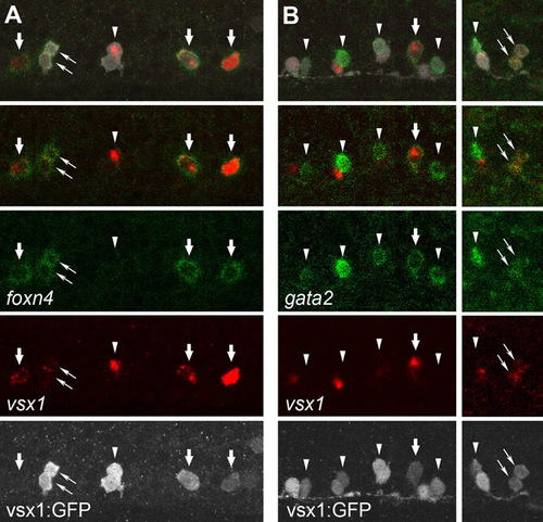
Expression profile of foxn4 and gata2 mRNA in the V2 neuron lineage. (A) Triple staining of foxn4 mRNA (green), vsx1 mRNA (red) and GFP protein (white) in a Tgvsx1:GFP embryo at 18 hpf. The top panel shows a merged image of three signals. The second panel from the top shows a merged image of the green and red channels. foxn4 is expressed in single vsx1-positive cells (thick arrows), which are likely to be p2 intermediate progenitors. The left-most cell that is positive for both foxn4 and vsx1 does not have detectable levels of GFP expression. The most likely explanation for this observation is that the cell is a very early stage p2 intermediate progenitor (accumulation of GFP has not yet reached at detectable level). foxn4-expressing cells can also be detected in a pair of vsx1-GFP cells (thin arrows), which are probably the progeny of a single progenitor. However, foxn4 expression can also be absent in both cells of a vsx1-GFP pair (arrowhead). This observation suggests that both of the cells have lost foxn4 expression. Thus, foxn4 mRNA expression in the V2 neuron lineage may proceed as follows: foxn4 expression is first upregulated in a p2 intermediate progenitor cell. After division, foxn4 is transiently present in both of the progeny. Subsequently, both cells lose foxn4 expression. It should be noted that the disappearance of foxn4 in the V2 progeny occurs earlier than that of vsx1. In the pair of cells marked by the arrowhead, vsx1 mRNA is still present in one of the cells (the vsx1-positive cell would probably differentiate into V2a neuron; see Fig. S1. (B) Triple staining of gata2 mRNA (green), vsx1 mRNA (red) and GFP protein (white) in a Tgvsx1:GFP embryo at 20 hpf. gata2 expression is expressed in single vsx1-positive cells (thick arrow) or in pairs of vsx1-GFP cells (thin arrows). In many of the paired cells, gata2 expression is detected only in one of the paired cells (arrowheads). gata2-negative vsx1-GFP cells usually express vsx1. Based on these observations, gata2 mRNA expression in the V2 neuron lineage may occur as follows: gata2 expression is first upregulated in p2 intermediate progenitors and after division is temporarily present in both of the progenies. One of the cells then loses gata2 expression (but continues to be positive for vsx1), and will differentiate into a V2a neuron. The other cell that continues to express gata2 will differentiate into a V2b neuron.
|
