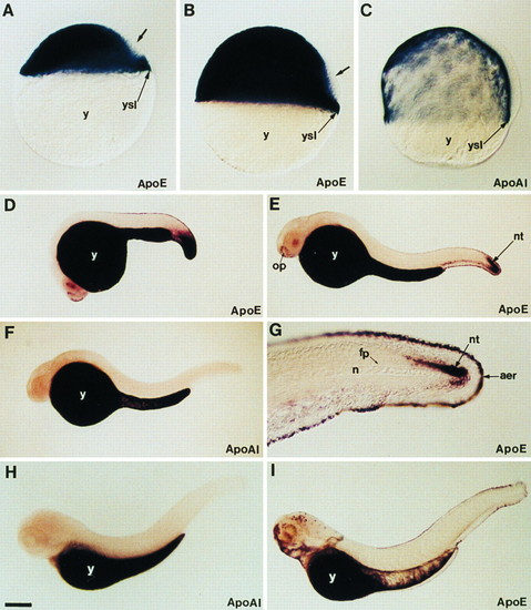
Expression pattern of apoE and apoA-I genes during embryonic and early larval development of zebrafish. (A and B) ApoE expression at 30% epiboly and shield stages, respectively. (C) ApoA-I expression at 70% epiboly. Both genes are expressed in the yolk syncytial layer (ysl) at these early stages while the yolk cell (y) does not contain any transcripts. The dorsal domain, which does not express apoE, is indicated by an arrow. (D) At 20 hr of development, apoE transcripts are located in the yolk syncytial layer and in the caudal region. (E) At 24 hr, apoE transcripts are detected in the yolk syncytial layer that covers the yolk cell in cells surrounding the olfactory placodes (op), in the ventral caudal mesoderm, and in the caudal part of the neural tube (nt). (G) High magnification of the tail region of a 24-hr-old embryo showing apoE transcripts in the apical ectodermal ridge (aer) of the fin and in caudal neural tube. Notocord (n) and floor plate (fp) are unlabeled. At 24 hr (F) and 3 days (H) of development, the expression of apoA-I gene is restricted to the yolk syncytial layer, while a new domain of apoE gene expression appears at 3 days in the head region, in the facial ectoderm, and in some cells of the retina and brain (I). Embryos were mounted in Permount (A-C) or in 100% glycerol and viewed with differential interference contrast optics (A-C and G) or with a dissecting microscope (D-F, H, and I). [Bar = 100 μm (A-C); 200 μm (D-F, H, and I); 50 μm (G).]
|
