
FIGURE
Fig. 2
Fig. 2
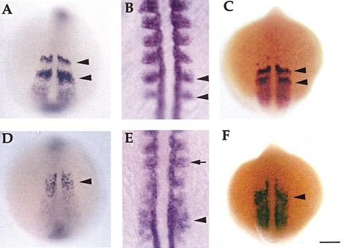
Somite and myotome gene expression is perturbed in desb420 mutants. Dorsal views (anterior to the top) of whole-mount RNA in situ hybridization of her1 (A, D; 13 h), myoD (B, E; 15 h), and mespa (C, F; 13 h) in wild-type (A–C) and mutant (D–F) embryos. Black arrowheads denote bands of gene expression in wild-type embryos and the corresponding region in mutants. Arrow in (E) designates normal myoD expression in anterior somites. Scale bar, 150 μm (A, C, D, F), 50 μm (B, E). |
Expression Data
Expression Detail
Antibody Labeling
Phenotype Data
| Fish: | |
|---|---|
| Observed In: | |
| Stage Range: | 5-9 somites to 10-13 somites |
Phenotype Detail
Acknowledgments
This image is the copyrighted work of the attributed author or publisher, and
ZFIN has permission only to display this image to its users.
Additional permissions should be obtained from the applicable author or publisher of the image.
Reprinted from Developmental Biology, 237(2), Gray, M., Moens, C.B., Amacher, S.L., Eisen, J.S., and Beattie, C.E., Zebrafish deadly seven functions in neurogenesis, 306-323, Copyright (2001) with permission from Elsevier. Full text @ Dev. Biol.