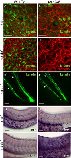
psoriasis mutants fail to express differentiation-specific keratins. A-D: Keratin expression in epidermal cells. Wild-type epidermal cells express and localize keratins basally at 3.5 (A) and 4.5 (C) days postfertilization (dpf), whereas psoriasis mutant epidermal cells have greatly decreased keratin expression at 3.5 (B) and 4.5 (D) dpf. The round green cells in B are unidentified, but are likely to be neuromast precursors based on regular spacing along the anterior-posterior axis in the position of the future lateral line. Cadherin staining (red) marks epidermal cell membranes. E,F: Keratin expression along the axis of the tail. Arrowheads indicate hypochord, and arrows indicate neural tube, respectively. Keratin expression is normal in this region of wild-type (E) and psoriasis mutants (F). E and F are lateral views with anterior to the upper left. G-J: In situ hybridizations for krt4. Wild-type embryos express krt4 at 48 hours postfertilization (hpf; G) and at lower levels at 3.5 dpf (I). psoriasis mutants express krt4 at 48 hpf similar to wild-type embryos (H), but exhibit increased krt4 expression at 3.5 dpf (J). Embryos in all these panels were stained for the same length of time. Anterior is to the left and dorsal is up in G-J. Scale bars = 25 μm in E,F, 50 μm in G-J.
|
