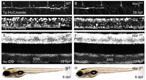
The kbpst23 mutation disrupts axonal development. (A,B) Lateral views of PLLn axons in live embryos bearing the Tg(HuC:kaede) transgene at 28 hpf. Axons of the wild type (A) have advanced further posterior than those in the kbpst23 mutant (B). The images show the segment of the nerve corresponding to about somites 6-12. Anterior is towards the left and dorsal towards the top. (C,D) Lateral views of spinal cord axons in live embryos bearing the Tg(HuC:kaede) transgene at 30 hpf. The arrowheads indicate the posterior-most axons in the ventral spinal cord tract in the wild type (C) and the kbpst23 mutant (D). The images show the segment of the spinal cord corresponding to about somites 14-20. Anterior is towards the left and dorsal towards the top. (E,F) Lateral views of embryos at 72 hpf showing neurons labeled with anti-HuC/D in the spinal cord (SC) and enteric nervous system (ENS) of a wild-type (E) and kbpst23 mutant (F). There are no discernable differences in neuronal organization between kbpst23 mutants and siblings. (G,H) Lateral views of live larvae at 6 dpf show no obvious morphological differences between wild-type (G) and kbpst23 mutant (H) larvae. The genotypes of all specimens were determined after photography (see Materials and methods).
|
