
Fig. 1
- ID
- ZDB-FIG-071204-25
- Publication
- Li et al., 2007 - Redundant activities of Tfap2a and Tfap2c are required for neural crest induction and development of other non-neural ectoderm derivatives in zebrafish embryos
- Other Figures
- All Figure Page
- Back to All Figure Page
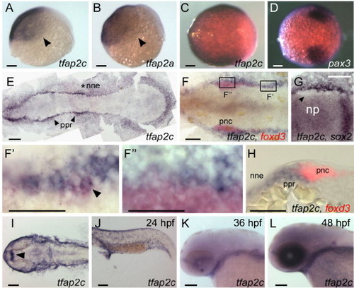
Comparison of tfap2a and tfap2c expression at embryonic stages. (A, B) Lateral views, animal pole oriented up and dorsal side to the right, of panel A, tfap2c and panel B, tfap2a expression in ventral and lateral ectoderm at 6 hpf. Arrowheads point to the limit of expression, which is similar or identical for the genes. (C, D) Animal pole views of panel C, tfap2c and panel D, pax3 expression at 8.5 hpf. Expression of pax3 marks prospective neural crest (Lewis et al., 2004); tfap2c expression partially or entirely overlaps it. (E–G) Dorsal views of flat-mounted embryos at 11 hpf, anterior to the left. (E) tfap2c expression is detected in non-neural ectoderm (nne, asterisk), with increased levels immediately adjacent to the rostral neural plate, in the pre-placodal region (ppr, arrowheads). (F) Double RNA in situ hybridization with foxd3 (red) and tfap2c (blue) probes. More caudal cells expressing foxd3, which are recently specified pre-migratory neural crest cells (pnc), also express tfap2c (shown at higher magnification in panel F′, arrowhead indicates double-labeled cell). By contrast, at more rostral levels, tfap2c is expressed in ppr but is excluded from the majority of foxd3 expressing cells (shown at higher magnification in panel F″). (G) An embryo labeled with tfap2c and sox2, a pan-neural plate marker (Okuda et al., 2006). tfap2c expression is absent from cells immediately lateral to the neural plate, which is the pnc domain (arrowhead). (H) A transverse section of an embryo labeled as in panel F. tfap2c (blue) is expressed at high level in the ppr and nne, lateral to foxd3 expression (red) in pnc. (I–L) Dorsal and lateral views of embryos at the indicated ages processed to reveal tfap2c. (I) In the head, and panel J, trunk at 24 hpf, expression of tfap2c is detected in surface ectoderm and diencephalon (arrowhead). (K) At 36 hpf, tfap2c expression is reduced in surface ectoderm; (L) at 48 hpf, high level tfap2c expression is detected in the eye. Scale bars: (A–G, I–L), 100 μm; (F′–H), 50 μm. |
| Genes: | |
|---|---|
| Fish: | |
| Anatomical Terms: | |
| Stage Range: | Shield to Long-pec |
Reprinted from Developmental Biology, 304(1), Li, W., and Cornell, R.A., Redundant activities of Tfap2a and Tfap2c are required for neural crest induction and development of other non-neural ectoderm derivatives in zebrafish embryos, 338-354, Copyright (2007) with permission from Elsevier. Full text @ Dev. Biol.