
Fig. 2
- ID
- ZDB-FIG-070802-29
- Publication
- Kok et al., 2007 - The role of the SPT6 chromatin remodeling factor in zebrafish embryogenesis
- Other Figures
- All Figure Page
- Back to All Figure Page
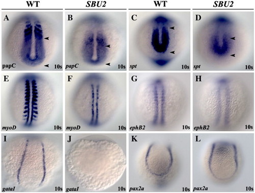
SBU2 mutants have mesoderm defects. Whole mount RNA in situ hybridization of mesodermal markers in wild-type and SBU2 mutants. All views are dorsal; anterior to the top. The expression domains of presomitic mesoderm markers papC and spt are reduced in SBU2 mutants (A–D). A–P patterning of the somites in SBU2 embryos is also affected; myoD expression at the posterior half of the somites is lost while it is reduced at adaxial cells (E and F), ephB2 at the anterior half of the somites is reduced and more diffused (G and H). gataI expression is diminished in SBU2 mutants (I and J) indicating that lateral plate mesoderm development is highly reduced. There is no apparent difference in pax2a expression between wild-type and SBU2 mutants (K and L); SBU2 mutants have no defect in intermediate mesoderm formation. The most anterior and posterior papC and spt expression is marked by arrowheads. |
| Genes: | |
|---|---|
| Fish: | |
| Anatomical Terms: | |
| Stage: | 10-13 somites |
Reprinted from Developmental Biology, 307(2), Kok, F.O., Oster, E., Mentzer, L., Hsieh, J.C., Henry, C.A., and Sirotkin, H.I., The role of the SPT6 chromatin remodeling factor in zebrafish embryogenesis, 214-226, Copyright (2007) with permission from Elsevier. Full text @ Dev. Biol.