FIGURE

Fig. 8

ID
ZDB-FIG-070802-26
Publication
Plaster et al., 2007 - REREa/Atrophin-2 interacts with histone deacetylase and Fgf8 signaling to regulate multiple processes of zebrafish development
Other Figures
All Figure Page
Back to All Figure Page
Fig. 8

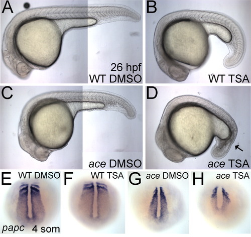
Histone deacetylase (HDAC) activity is required for proper Fgf signaling during development of the posterior mesoderm. A-D: Lateral views of live 26 hours postfertilization (hpf) wild-type (A,B) or ace (C,D) embryos, treated with either dimethylsulfoxide (DMSO) control medium (A,C) or 200 nM Trichostatin A (TSA, B,D) are shown. TSA-treated embryos are slightly delayed. The black arrow in D marks severely disrupted tail mesoderm, resembling the bab;ace phenotype at 24 hpf. E-H: Whole-mount in situ hybridizations (dorsal views on the tail bud) of papc at the four-somite stage in wild-type embryos (E,F) or ace (G,H) treated with DMSO control medium (E,G) or 200 nM TSA (F,H) from 30% epiboly.

Expression Data
Gene:
Fish:
Condition:
Anatomical Term:
Stage: 1-4 somites

Expression Detail
Antibody Labeling
Phenotype Data
Fish:
Condition:
Observed In:
Stage Range: 1-4 somites to Prim-5

Phenotype Detail
Acknowledgments
This image is the copyrighted work of the attributed author or publisher, and ZFIN has permission only to display this image to its users. Additional permissions should be obtained from the applicable author or publisher of the image. Full text @ Dev. Dyn.