
Fig. 7
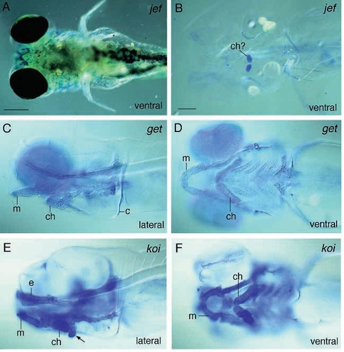
Photomicrographs of several members of the hammerhead-like group stained with Alcian blue. Mutant jellyfish embryos (A,B) can be identified by a severe lack of tissue anterior to the eyes (A, ventral view) and strongly reduced or loss of cartilage. Two elements are always present at the level of the ceratohyals (B, ventral view). In geist (ti240) mutant larvae (C,D) the elements hardly stain with Alcian blue and consist of less organized chondrocytes. Tumor-like outgrowths of chondrocytes are characteristic for knorrig mutant larvae (E,F). From a lateral view (E), additional chondrocytes can be seen, particularly ventral to the ceratohyal (arrow). A ventral view (F) reveals that none of the elements possesses well defined edges. Scale bars: 200 μm (A) and 100 μm (B-F). |