
Fig. 7
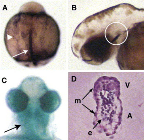
neuropilin1A is expressed in LPM and morphant phenotypes are similar to those of sema3D morphants. (A) At 8 SS, nrp1A RNA is strongly expressed in LPM (shown in arrowhead) and in the notochord (shown in arrow), not in neural crest. Dorsal view of embryo, cranial to the top. (B) rag1 expression in the thymus (white circle) was reduced in morphants. (C) Alcian blue staining showed defective facial and pharyngeal cartilage development (arrow, ventral view). (D) nrp1A morphants have dysmorphic hearts with smaller ventricle (V), smaller atrium (A) and thickened myocardium (m). Endocardium was present (e) (4 dpf, lateral section, H&E stain). |
| Genes: | |
|---|---|
| Fish: | |
| Knockdown Reagent: | |
| Anatomical Terms: | |
| Stage Range: | 5-9 somites to Day 4 |
Reprinted from Developmental Biology, 298(1), Sato, M., Tsai, H.J., and Yost, H.J., Semaphorin3D regulates invasion of cardiac neural crest cells into the primary heart field, 12-21, Copyright (2006) with permission from Elsevier. Full text @ Dev. Biol.