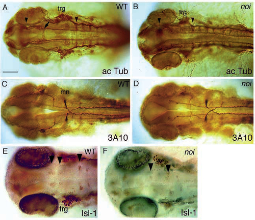
Overall organisation of the CNS in noi mutant embryos. Shown are dorsal views of wild type and mutant embryos stained with antibodies to reveal axonal organisation and formation of neuronal nuclei. (A,B) Anti-acetylated-tubulin staining in 30 h wild-type and mutant noity22b embryos. Arrowheads point to the position of the tract of the posterior commissure, and to the first hindbrain commissure. Notice the reduced distance. The arrow in A points to the ventral tegmental commissure that is absent in the mutant noi embryo in B. (C,D) mAb 3A10 staining in 30 h wild-type and mutant noith44 embryos. The Mauthner neuron (mn) and the other T reticular interneurons stained by this antibody develop normally, indicating that hindbrain development is largely normal in noi embryos. (E,F) Isl staining of 32 h wild-type and mutant noith44 embryos. Arrowheads point to bilateral pairs of clustered neuronal nuclei stained by this antibody in the midbrain and hindbrain (see text). Whereas two clusters are seen in the midbrain of the wild-type embryo in E (probably corresponding to the nucleus of the medial longitudinal fascicle), only one such pair of reduced size is seen in the mutant. Notice the reduced distance to the rhombomere 2 cluster. A group of 2-3 nuclei in rhombomere 1 is also unaffected in the mutant, but not in focus. trg, trigeminal ganglion. Scale bar, 100 µm (A-D), 75 µm (E, F).
|
