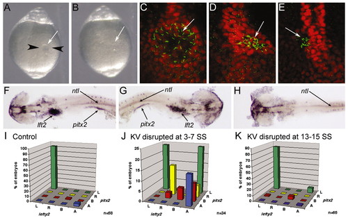
KV is essential for LR development during early somitogenesis. (A) Image of a live 6 SS embryo prior to disruption of KV. Black arrowheads represent the path of the needles during KV disruption and the white arrow indicates KV. (B) Image of the same embryo in A after disruption of KV. White arrow indicates where KV was previously located. (A,B) Dorsal views of the tail. (C-E) Control embryo (C) and KV disrupted embryos (D,E) stained with anti-acetylated tubulin (green) and ntl (red) antibodies at 10 SS. Arrows indicate KV cilia. (F-H) In situ hybridization analysis of lft2, pitx2 and ntl at 20-25 SS. (F) A control embryo showing normal left-lateral gene expression. KV disrupted embryos often showed either reversed (G) or an absence (H) of lateral gene expression. Arrows indicate lft2 staining in the heart primordia, pitx2 staining in the lateral mesoderm and ntl staining in the notochord. Yolk was dissected away for visualization of lateral staining. (F-H) Dorsal views with the anterior towards the left in F,H and to the right in G. (I-K) lft2 expression in the heart primordia plotted against pitx2 expression in the lateral plate mesoderm in control (I), KV-disrupted at 3-7 SS (J) and KV-disrupted at 13-15 SS (K) embryos. L, left; R, right; B, bilateral; A, absent. n, number of embryos examined.
|
